Division of Transdimensional Life Imaging, Institute for Open and Transdisciplinary Research Initiatives, Osaka University
![]()
RESEARCH
Research
1. Development of trans-scale imaging system AMATERAS
We have developed the first trans-scale imaging system AMATERAS (AMATERAS1.0), which is the key technology for research in our division [1]. The fluorescence imaging system was constructed using an ultra-multipixel CMOS camera and a telecentric macro lens for machine vision, instead of using the lenses and cameras of a biological microscope, to establish an ultra large field-of-view (FOV) cell imaging technique. Fluorescence imaging of the dynamics and function of individual cells can be observed in an FOV of 14.6mm x 10.1mm, which is the world's largest field of view as an optical imaging system with cellular resolution. With this new imaging system, 100,000~1,000,000 cells can be observed simultaneously (Fig. 1a). Imaging of a section of a mouse brain has shown that individual cells of the entire brain can be observed simultaneously (Fig. 1b). We are promoting applications to research that will lead to a better understanding of the mechanisms of multicellular systems, such as statistical analysis of the diversity and inhomogeneity of cellular states.
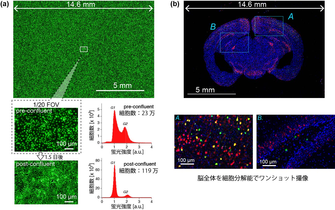
Fig. 1: Observation of cell populations by AMATERAS. (a) Fluorescent image of epithelial cells. 230,000 cells were observed simultaneously in one-shot imaging (top, pre-confluent). Furthermore, 1.5 days later, the cells proliferated and more than 1 million cells (1.19 million) could be observed simultaneously (post-confluent). Image analysis enabled analysis of cell cycle distribution (bottom right). (b) Fluorescent image of a mouse brain section. Individual cells in the whole FOV were observed by one-shot imaging.
2. Application of AMATERAS: Detection of leader cells triggering spatial pattern formation in social amoebae
In collaboration with the laboratory of Prof. Horikawa at Tokushima University, we have observed spatial pattern formation in the social amoeba, Dictyostelium discoideum, by AMATERAS to elucidate the pattern formation mechanism and detect leader cells [1,2]. These cells have the property of aggregating to form slug-like multicellular bodies when nutritional resources are depleted. In the assembly process, it is known that cAMP molecules generated inside the cells are released to the outside of the cells, which are repeatedly accepted by other cells, thereby promoting cell assembly. A complex of two fluorescent proteins (Flamindo2 and mRFP) was used as a cAMP indicator, and the spatial distribution of intracellular cAMP concentration was visualized by their fluorescence intensity ratio (mRFP/Flamindo2) dependent on cAMP concentration (Fig. 2A). More than 230,000 cells were observed in the entire field of view. Compared to the uniform spatial distribution in the initial stage (Fig. 2B upper), the spiral wave propagated 2.5 hours later (Fig. 2B middle), and finally a multicellular body was constructed (Fig. 2B lower), allowing simultaneous observation of the overall spatial pattern and individual cell dynamics. In particular, we focused on the mechanism of spiral wave generation, and by analyzing images at the cellular granular level, we discovered minority cells, which we called "leader cells," that cause the generation of spiral waves, and clarified the mechanism by which leaders generate spiral waves. We found that cells that behave as leaders exist at a rate of about 130 cells per 100,000 cells (0.13%) [2].
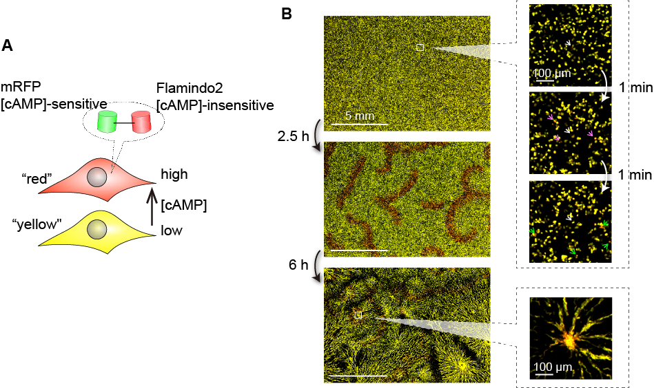
Figure 2: Observations of the social amoeba Dictyosterium Discoideum. (A) Fluorescent protein sensor for cAMP visualization. (B) Results of two-wavelength time-lapse measurement. Left: spatial pattern image of the entire field of view; right: dynamics of individual cells in local regions.
References
[1] "Exploring rare cellular activity in more than one million cells by
a transscale scope,"
Scientific Reports 11, Article number: 16539 (2021).
https://doi.org/10.1038/s41598-021-95930-7
[2] "Cellular logics bringing the symmetry breaking in spiral nucleation
revealed by trans-scale imaging,"
bioRxiv (2020).
https://doi.org/10.1101/2020.06.29.176891
バナースペース
Division of Transdimensional Life Imaging, Institute for Open and Transdisciplinary Research Initiatives, Osaka University
Yamadaoka 2-1, Suita, Osaka 565-0871
TEL +81-6-6105-6263